CORONARY CT: Non-Invasive Test in the Detection of Coronary Artery Disease

By Anush Parikh, M.D
Coronary artery disease (CAD), heart disease, atherosclerotic disease, cardiovascular disease (CVD); the number one killer of Americans goes by many names. Despite the various nomenclatures, the underlying commonality is blockage of arteries of the heart which can lead to heart dysfunction/stoppage (heart attack or cardiac arrest).
CAD is the buildup of cholesterol/fatty and calcified deposits (plaque) within the arteries of the heart. This overtime leads to narrowing of the blood vessel which can eventually become so severely narrowed that adequate blood flow/oxygen cannot be delivered to the heart muscles. This can produce angina (chest pain with nausea, fatigue, shortness of breath, sweating among other symptoms). Alternatively, the plaque can rupture and lead to immediate blockage of the arteries and subsequent sudden cardiac arrest. There are many risk factors for CAD, with the major ones being increasing age, male gender, genetics, smoking, high cholesterol, high blood pressure, low physical activity, obesity, diabetes, stress, and excessive alcohol consumption.
According to the 2009 statistics from the AHA (American Heart Association) one in three female adults has some form of CVD. Since 1984, the number of CVD deaths for women has exceeded the deaths for men. The AHA statistics state that in 2009, CVD was the cause of death in 401,495 women. Women represented 51% of deaths from cardiovascular disease. Approximately 66 million women alive today have coronary heart disease.
The primary test to image the arteries of the heart has been an invasive procedure
known as cardiac catheterization which involves puncturing the femoral artery followed by catheters and wires which are guided from the groin to the heart and evaluated by x-ray. MDI has the technology to accurately and non-invasively image the arteries of the heart with our 64 detector/slice CT scanner (CAT scan) which is scheduled to be upgraded to an even more powerful and higher resolution state-of-the-art 128 detector/slice CT scanner in 2016. This test is known as Coronary CT Angiography.
The technologist will place an IV and monitor your heart rate and may give you a low dose medication to control it (beta blocker). Iodine contrast material is pushed through the IV and the 64/128 slice CT scanner obtains extremely thin (0.5 mm) images through the heart. These images capture details of the moving heart and its arteries which allow us to construct high resolution 3-D images to determine if there is any fatty/cholesterol or calcified deposits (plaque) narrowing the arteries.
A study performed at the Cornell Medical Center in New York City found that the non-invasive Coronary CT Angiography study has a very high negative predictive value (97-99%) in patients with chest pain who are electively referred for invasive coronary angiography. The higher the negative predictive value, the more effective a test is at excluding disease. Thus, Coronary CT Angiography is a highly effective noninvasive alternative to exclude coronary artery narrowing/stenosis.
Speak to your doctor to see if this test may benefit you or call us for more information regarding Coronary CT angiography or any CAT scan/radiologic procedure.
TESTIMONIALS:
“My father had a heart attack at a young age. I am 58 years old and had chest pain on and off for a year with inconclusive tests. I had the coronary CT and it showed a severe blockage. I was one of the lucky few who was diagnosed early enough before a heart attack.” – Lisa M.
“I had vague chest discomfort and have smoked for 30 years. I went to MDI where the incredibly helpful staff walked me through the coronary CT which found a 70% blockage and saved my life.” Jen D.
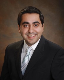
Anush Parikh, M.D. is with Mid-Delaware Imaging in Dover, DE. Dr. Parikh was raised in Dover and educated through the Holy Cross, Caesar Rodney and St. Andrew’s school systems. He attended New York University for undergraduate schooling and majored in Anthropology. Dr. Parikh received his M.D. at Syracuse-Upstate Medical University where he achieved the medical academic honor of Alpha Omega Alpha. He moved back to NYC where he completed his Internship at St. Vincent’s Catholic Medical Center. This was followed by a residency in Diagnostic Radiology at Beth Israel Medical Center and a mini-fellowship in cardiac/coronary CT.


